
-
Topics
subnavigation
Topics
Electromagnetic fields
- What are electromagnetic fields?
- Static and low-frequency fields
- Radiation protection relating to the expansion of the national grid
- High-frequency fields
- Radiation protection in mobile communication
Optical radiation
Ionising radiation
- What is ionising radiation?
- Radioactivity in the environment
- Applications in medicine
- Applications in daily life and in technology
- Effects
- What are the effects of radiation?
- Effects of selected radioactive materials
- Consequences of a radiation accident
- Cancer and leukaemia
- Genetic radiation effects
- Individual radiosensitivity
- Epidemiology of radiation-induced diseases
- Ionising radiation: positive effects?
- Risk estimation and assessment
- Radiation protection
- Nuclear accident management
- Service offers
-
The BfS
subnavigation
The BfS
- About us
- Science and research
- Laws and regulations
- BfS Topics in the Bundestag
- Links

X-ray diagnostics – the procedure

In X-ray diagnostics, a distinction is made between three techniques:
- radiography,
- fluoroscopy,
- computed tomography (CT) and other tomographic techniques.
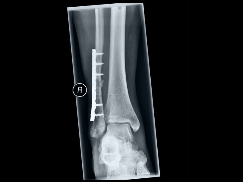
1. Radiography
The most frequently used technique is the conventional radiography which is associated with a comparatively low radiation exposure. The part of the body to be examined is exposed to X-rays for a fraction of a second and the radiation penetrating the body is visualised using a digital detector or – in rare cases – still a film-screen system. The reproduction of dense structures such as bone is light, whereas that of less dense tissues such as fatty tissue is dark. Structures of mean density such as soft tissues (e.g. cartilage), are represented in different shades of grey.

 Fluoroscopy
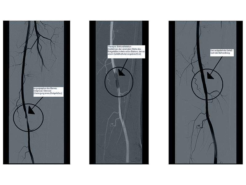
2. Fluoroscopy
Examinations of movement sequences (such as the act of swallowing, or the heartbeat) or more precise assessments of overlapping structures (e.g. of the gastro-intestinal tract) sometimes require additional fluoroscopy. This technique involves soft X-radiation penetrating the body to produce a series of pictures represented on a luminescent screen. By means of electronic image intensification these pictures are subsequently transferred to a monitor for visual examination.
Fluoroscopy also includes angiography. This technique involves a contrast medium administered to the patient in order to reproduce vessels which normally cannot be visualised. Angiography may be accompanied by so-called interventional measures (serving therapy instead of diagnosis) such as the widening of narrowed or blocked blood vessels.
The radiation dose to the patient associated with fluoroscopy may be well above that occurring during standard radiography.
")
 Computed tomography (CT)
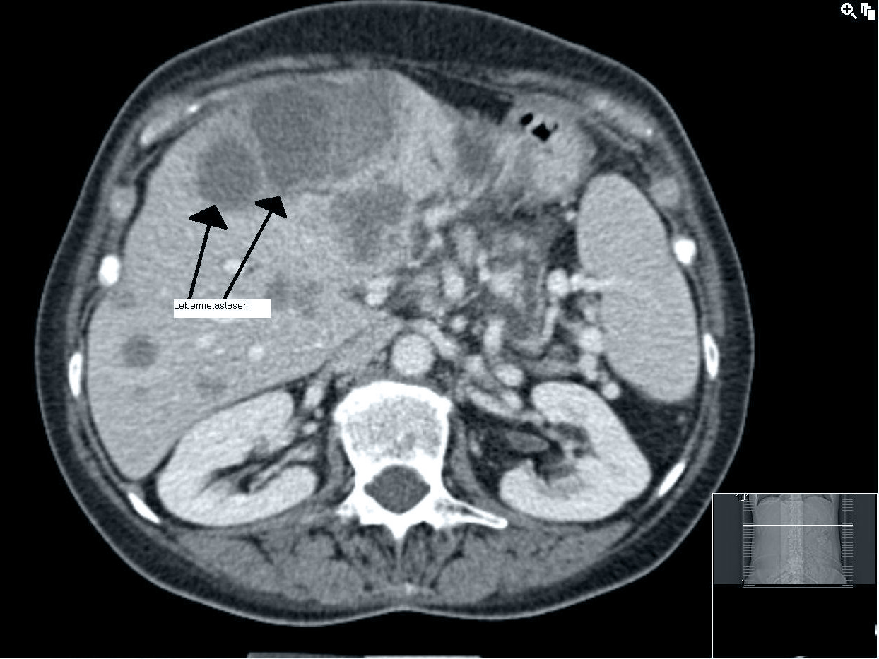
3. Computed tomography (CT) and other tomographic techniques
CT is a procedure of X-ray diagnostics creating cross-sectional pictures of the body. During CT an X-ray machine, consisting of an X-ray tube assembly and a radiation detector on the opposite site, continuously rotates around the body, following a circular or spiral path to obtain a multiplicity of radiographs from different directions (projections). A computer program is used to transform these projection images into cross-sectional non-overlapping pictures of high-contrast displaying tissues of different density in well distinguishable shades of grey.
The radiation dose to the patient caused by CT is relatively high as compared to radiography.
In recent times, dental cone beam CT and digital breast tomosynthesis (DBT) were introduced as additional sectional imaging techniques. Dental cone beam CT is mainly used in dentistry, orthodontics, and otolaryngology to reduce the problem of superposition, e.g. for a more precise treatment planning. DBT can e.g. be used if a suspicious finding after conventional mammography needs further investigations, and alternative examination techniques have been unrewarding.
State of 2018.04.09