
-
Topics
subnavigation
Topics
Electromagnetic fields
- What are electromagnetic fields?
- Static and low-frequency fields
- Radiation protection relating to the expansion of the national grid
- High-frequency fields
- Radiation protection in mobile communication
Optical radiation
Ionising radiation
- What is ionising radiation?
- Radioactivity in the environment
- Applications in medicine
- Applications in daily life and in technology
- Effects
- What are the effects of radiation?
- Effects of selected radioactive materials
- Consequences of a radiation accident
- Cancer and leukaemia
- Genetic radiation effects
- Individual radiosensitivity
- Epidemiology of radiation-induced diseases
- Ionising radiation: positive effects?
- Risk estimation and assessment
- Radiation protection
- Nuclear accident management
- Service offers
-
The BfS
subnavigation
The BfS
- About us
- Science and research
- Laws and regulations
- BfS Topics in the Bundestag
- Links
Influence of static magnetic fields on reproduction and development
- From 2008 to 2011, scientists from the University of Duisburg-Essen, Germany, conducted studies on laboratory animals (mice) to answer the question of whether, and if so, how, strong static magnetic fields influence the reproduction (fertility, pregnancy) of mammals and the development of embryos as well as the further physical development of the offspring.
- The fertility of male and the course of pregnancy of female mice were not affected by repeated strong magnetic field exposure.
- The offspring exposed during embryonic development did not show any health relevant effects of exposure. However, a slightly delayed development in terms of weight and opening of the eyes was observed as compared to controls. It is possible that the developmental delay was induced by stress during exposure.
During the last few decades, magnetic resonance imaging has been applied increasingly also in pregnant women and for the purpose of gynaecologic diagnosis. In order to improve image quality, higher-performance scanners have been developed, resulting in enhanced flux density, which is why a review of biological safety is required.
Within the scope of the research project “Effects of repeated exposure to strong static magnetic fields from MRI on the end points reproduction and development in an animal model” ("Auswirkungen wiederholter Exposition mit starken statischen Magnetfeldern des MRT auf die Endpunkte Fortpflanzung und Entwicklung im Tiermodell"), lasting from 2008 to 2011, scientists from the University of Duisburg-Essen, Germany, conducted studies on laboratory animals to answer the question of whether, and if so, how, strong static magnetic fields influence the development of sperms in male adult mice, and the pregnancy and development of embryos in female mice. In addition, fertility of male and female mice exposed to static magnetic fields daily throughout the entire period of embryonic development was studied.
The results are of particular importance for the safety of both pregnant patients and medical staff.

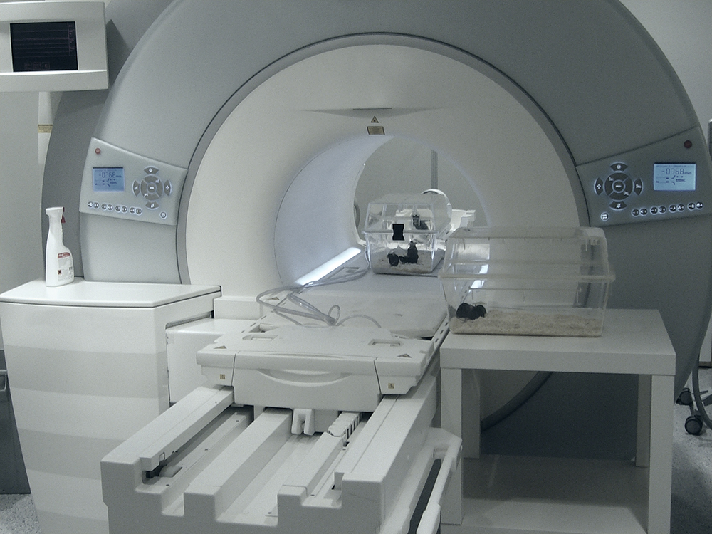 Mice in and in front of the scanner
Source: University Duisburg-Essen
Mice in and in front of the scanner
Source: University Duisburg-Essen
Method
Male and pregnant female mice were exposed at the isocentre, and at the entrance of a 1.5 Tesla and a 7 Tesla MRI Scanner 75 minutes a day for 21 days, or sham-exposed for control (0 Tesla).
Exposed males and females
Fertility of exposed males was studied by reference to sperm activity and morphology as well as to the testicular state and was found to be not affected. In females, magnetic field exposure turned out to have no influence on pregnancy rate, duration, litter size and gender distribution in the offspring. No resorptions (loss of embryos) were observed.
Offspring exposed during embryonic development
No malformations (funicular hernia, cerebral hernia, malformations of limbs) were observed among the young animals exposed during embryonic development. Eye malformations occurred in very rare cases and showed no correlation with magnetic field exposure, revealing the same frequency in unexposed and exposed animals.
Compared to controls most of the groups exposed during embryonic development were found to exhibit slightly delayed development in terms of weight and opening of their eyes, whereby the animals exposed at seven Tesla were least affected. All values were in the normal physiological range and the animals were healthy.

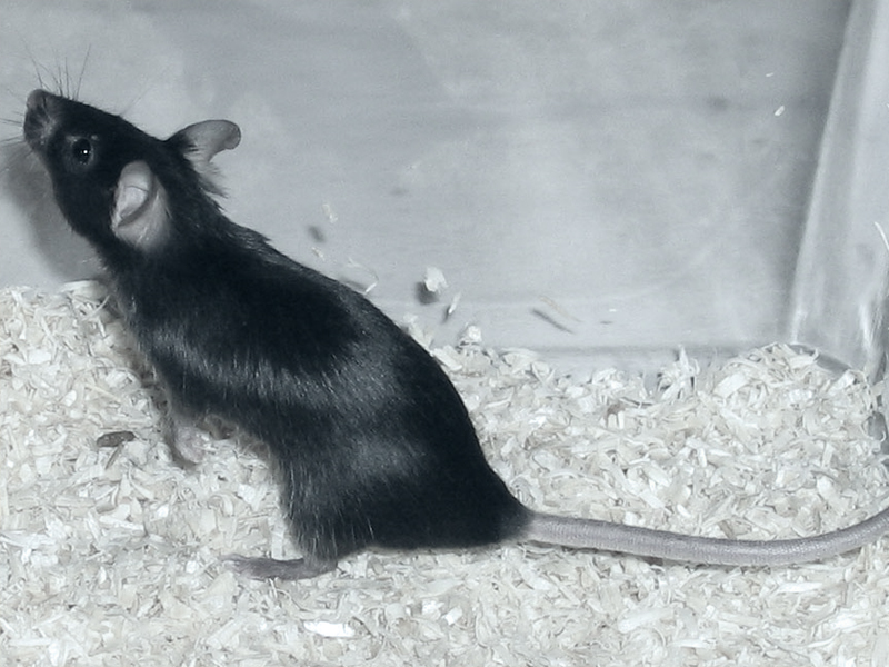 C57BL/6 Mouse
Source: Zentralinstitut für seelische Gesundheit Mannheim
C57BL/6 Mouse
Source: Zentralinstitut für seelische Gesundheit Mannheim
Offspring fertility
Magnetic field exposure was found to have no influence on testicles and sperms in the young males exposed during their embryonic development. When mated with unexposed females, these males were just as fertile as sham-exposed animals, as measured by the pregnancy rate.
Exposure of young females during their embryonic development was observed to have no influence on the pregnancy rate, number of embryos and resorption rate subsequent to mating with unexposed males. However, the females exposed to static magnetic fields during their embryonic development exhibited significantly lighter placentas, and at seven Tesla the embryos were found to be significantly lighter, too. Although the lower weight of embryos and placentas could be indicative of an adverse effect of exposure, all embryos were healthy and normally developed.
Discussion
The animals exhibited changes in behaviour during exposure in that they obviously moved less than unexposed animals, suggesting that they had perceived the fields. In the literature there are reports suggesting that rodents can perceive static magnetic fields and experience them as annoying. This could have caused stress which in turn might have resulted in the observed developmental delay.
It has been well established in the literature that stress affects embryonic development. The research project on cognitive performance demonstrated that exposure up to seven Tesla does not induce measurable stress in humans. The above mechanism of action therefore appears to be rather unlikely in women.
The statistical evaluation of the study was designed to ensure that even slight effects were preferably not overlooked. No correction for multiple tests was made. This has the advantage that potential adverse health effects are not missed, and therefore can be largely excluded. The disadvantage is that false-positive results may occur. Whether or not this is applicable for the developmental delay can be neither confirmed nor excluded.
The cognitive and emotional behaviour of the offspring exposed during their embryonic development was studied within the scope of a second project which revealed no delay in the development of the nervous system and behaviour.
Summary
The fertility of male and the course of pregnancy of female mice were not affected by repeated strong magnetic field exposition. The offspring exhibited a slightly delayed development in terms of weight and opening their eyes, as compared to controls. It is possible that the developmental delay was induced by stress during exposure.
State of 2018.07.27