
-
Topics
subnavigation
Topics
Electromagnetic fields
- What are electromagnetic fields?
- Static and low-frequency fields
- Radiation protection relating to the expansion of the national grid
- High-frequency fields
- Radiation protection in mobile communication
Optical radiation
Ionising radiation
- What is ionising radiation?
- Radioactivity in the environment
- Applications in medicine
- Applications in daily life and in technology
- Effects
- What are the effects of radiation?
- Effects of selected radioactive materials
- Consequences of a radiation accident
- Cancer and leukaemia
- Genetic radiation effects
- Individual radiosensitivity
- Epidemiology of radiation-induced diseases
- Ionising radiation: positive effects?
- Risk estimation and assessment
- Radiation protection
- Nuclear accident management
- Service offers
-
The BfS
subnavigation
The BfS
- About us
- Science and research
- Laws and regulations
- BfS Topics in the Bundestag
- Links

Magnetic Resonance Imaging

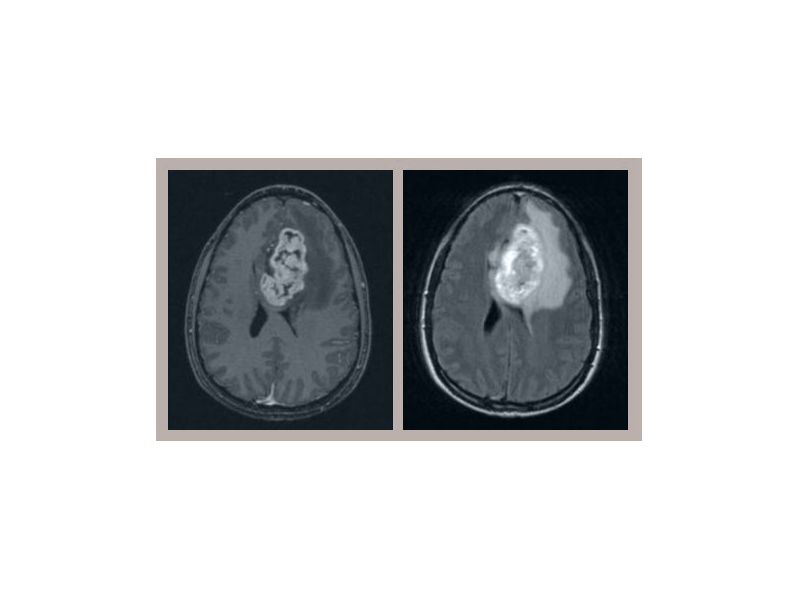 Magnetic resonance images of a patient with a brain tumour. As the images illustrate, the tissue contrast can be varied in magnetic resonance imaging.
Source: Quelle: DKFZ, Heidelberg
Magnetic resonance images of a patient with a brain tumour. As the images illustrate, the tissue contrast can be varied in magnetic resonance imaging.
Source: Quelle: DKFZ, Heidelberg
Magnetic resonance imaging (MRI) systems for clinical use have been available since the early 1980s. Due to the rapid technological development, MRI has been established as an indispensable tool for imaging diagnostics. In 2014, about 11 million examinations were carried out in Germany.
Advantages of MRI in medical diagnostics are that no ionising radiation is involved and the high soft-tissue contrast of the images (see figure). Furthermore, the tissue contrast in MRI - as opposed to computed tomography (CT) - can be varied over a wide range by selecting appropriate measurement parameters. In contrast, the latest generation of CT systems makes it possible to visualise anatomical structures with high spatial and temporal resolution which is particularly important for the diagnostics of moving organs.
MRI is used routinely and very successfully for a variety of issues related to various medical specialties. The clinical focus is on the diagnosis of oncological, inflammatory and degenerative diseases as well as of abnormalities and occlusions of blood vessels (e.g. diagnosis of infarction). For some of these purposes, there are no alternatives to MRI diagnostics.
Types of fields used in MRI
Three different fields are used in MRI:
- a strong static magnetic field generating a macroscopic nuclear magnetization,
- pulsed electromagnetic radiofrequency fields for excitation and preparation of the spin system as well as
- rapidly alternating magnetic fields with a linear spatial dependence (so-called magnetic gradient fields) for spatial encoding of the MRI signal.
In the following, the essential interaction mechanisms and biological effects of these three fields are briefly explained and safety recommendations are given. More detailed information is provided in the "Recommendations on the safe use of Magnetic Resonance Imaging (MRI) in medical diagnosis" by the German Commission on Radiological Protection (SSK) from 2002.
Biological effects and safety recommendations
Static magnetic fieldshow / hide
Although there is evidence for a multitude of different effects of static magnetic fields, particularly on macromolecules and flowing charges, studies published to date provide no indication of adverse health effects at whole body exposures of up to 4 T.
However, a substantial potential hazard in clinical routine, which cannot be prevented by complying with this reference value either, are metallic and especially ferromagnetic objects such as
- coins,
- scissors,
- and drip stands.
These objects are strongly accelerated in the stray field of the magnet and thus turn into dangerous projectiles.
This hazard can only be minimised by providing patients and possible escorts with detailed information. Furthermore, the operator of an MRI system is obliged to post warning signs alerting people to the danger.
Electromagnetic radiofrequency fieldsshow / hide
A fundamental biophysical effect of electromagnetic radiofrequency fields is the heating of tissue. The reason for this are electric currents induced in tissues, leading to power deposition and thus to heating due to electrical resistance of tissues. The energy absorbed by the body per mass and time is referred to as specific absorption rate (SAR, in watts per kilogram).
The relevant parameter to assess the physiological effect of radiofrequency fields is the temperature rise in tissues which does not only depend on local power absorption and exposure period, but also on the thermal conduction and tissue perfusion. The two last-mentioned factors lead to a quick temperature equilibrization in the case of regional heating of the body.
Investigations on humans have not revealed any adverse health effects from radiofrequency exposure of the torso and head yielding a maximum body temperature rise of 1 °C.
The SSK has recommended basic restrictions for temperature and limits for the specific absorption rate derived from these, compliance with which is ensured by complex monitoring systems during patient examinations.
Gradient fields show / hide
Rapidly alternating magnetic gradient fields may cause stimulation of peripheral muscles and nerves as well as stimulation of the heart muscle with the possible consequences ranging from extrasystoles to ventricular fibrillation. When using ultra-fast MRI techniques under experimental conditions, contractions occurred in chest area, shoulders, hips, buttocks and nose.
As it is not clear whether nerve or muscle cells are stimulated in these cases, such occurrences are generally referred to as magnetostimulation. From theoretical considerations and experimental observations, however, it is known that stimulations are only possible when the stimulus intensity exceeds a certain threshold and that cardiac thresholds are considerably higher (by a factor of about ten) than those required for the stimulation of peripheral nerves and muscles.
In the above-stated recommendation of the SSK, exposure limits are set which have to be complied with during patient examinations in order to avoid the stimulation of peripheral nerve and muscle cells. Compliance with these limits is ensured during patient examination performed at an approved MRI system by a control system.
In addition, considerable noise, which may require the use of hearing protection devices, can be generated by rapidly alternating gradient fields.
Further recommendations for protecting patients at risk
The SSK demands careful consideration of the indication for MRI and special monitoring during the examination in case of infants and toddlers as well as patients with impaired thermoregulation, severely disturbed blood circulatory, cardiac arrhythmias, poor general condition or known epilepsy. Special preventive measures are necessary also during pregnancy.
MR examinations of patients with implants or metallic inclusions are always associated with potential hazard even if the exposure limits stated above are complied with. This hazard can only be limited by carefully interviewing the patient, reviewing the patient's medical record and contacting producers or the attending physicians for further information. Prior to each examination of patients at risk, the risks and the expected diagnostic benefits have to be balanced carefully.
Patients with passive implants or metallic inclusionsshow / hide
Prior to the MRI examination of patients with passive implants such as
- vascular clips or clamps
- intravascular filters or stents,
- artificial heart valves,
- intravenous catheters or ports,
- orthopaedic prostheses,
- plates or screws as well as
- intrauterine coils
it has to be clarified whether these implants are ferromagnetic or whether they contain ferromagnetic components.
Evaluating the situation is more difficult in the case of metallic inclusions such as splinters or bullets, the materials of which are mostly unknown. In these cases, MRI examinations should not be carried out. This also applies to patients in whom critical tissue or organ structures are at risk due to potential dislocations of metallic inclusions or implants.
The risk potential is lower when inclusions or implants have grown in well and are not near critical structures. In such cases, MRI examinations are carried out in many places. However, these examinations should be limited to urgent medical indications.
Patients with active implantsshow / hide
MR examinations are contraindicated in patients with active implants unless otherwise specified on the patient implant card or by the attending physician.
In addition to the risks described above, the MRI system may modify or impair the operation of these devices and may thereby result in a hazard to the patient.
Important examples to be mentioned in this context are electric and mechanic components of pacemakers and infusion pumps. Furthermore, a range of active implants (such as cochlear implants) use small magnets for activation, programming or controlling functionality which means that an interaction between the implant and the fields used in MRI can cause the device to malfunction.
MRI during pregnancyshow / hide
MRI examinations during pregnancy should be performed only after a critical risk/benefit analysis and at the lowest possible exposure levels and shortest possible exposure time.
Especially during the first three months of pregnancy, particularly strict indication criteria have to be applied.
State of 2018.07.23